Today’s most advanced microscopes can reveal biology in stunning detail, from molecular machines operating inside cells to the development of an entire organism. But much of what drives life at the cellular level — proteins folding, cargo moving among organelles — occurs at scales and speeds that current state-of-the-art microscopy still can’t capture. Closing that gap has become one of the central bottlenecks in biological research — and in building the data that AI models need to predict how cells behave.
Biohub’s new Imaging Residency Program brings leading investigators from around the world and embeds them into its research teams to solve these problems together. So far, 18 scientists have been selected to work with Biohub teams in the areas of cryo-electron tomography, computational imaging, developmental systems mapping, immunological surveillance, and more. Under a collaborative research agreement, Imaging Residents are given a fixed amount of funding to work on a focused project.
“Biohub has ambitious goals, and to accomplish them, we want to bring the scientific community along with us and partner with experts to accelerate innovation,” says Stephani Otte, Biohub’s vice president of imaging science. “A major priority of our work in imaging is developing the technology to measure and map complex systems.”
Together, the Imaging Residents and members of their lab work closely with Biohub scientists on projects spanning a wide range of challenges, from camera hardware for fluorescence lifetime imaging microscopy (FLIM), to compression schemes for petascale microscopy data, to bioelectric mapping in developing zebrafish embryos. What began as a pilot last year has grown into an ongoing program that is already yielding significant technical advances, while building deep collaborations across institutions and disciplines.
Early successes
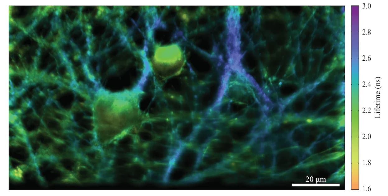
The residents are already expanding what imaging can capture, in time and space. Adam Bowman of the Salk Institute, one of the earliest Imaging Residents, has been working with Biohub scientist Shu-Hsien Sheu to develop a system that can capture nanosecond-scale time information. They built a next-generation 3D electro-optic version of FLIM (EO-FLIM), which allows scientists to capture a fluorescent molecule’s fluorescence lifetime – usually a few billionths of a second. Because that lifetime can change depending on the molecule’s environment, that enables the molecule to act as a biological sensor in response to conditions such as acidity, calcium concentration, electrical activity, or interactions between proteins.
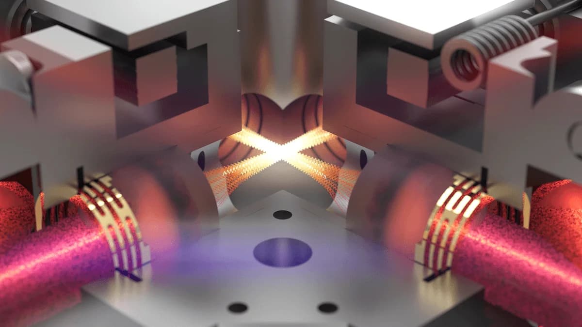
Together, Bowman and Sheu integrated the EO-FLIM into a single-objective light-sheet microscope at Biohub, creating the first microscope capable of very rapid FLIM in a 3D volume. Scientists can use the system to get quantitative information on fast processes that are otherwise hard to obtain, such as watching calcium propagate within a human neuron and measuring where, when, and how strong the signals are. “What this allows us to do is to make much more quantitative measurements in biology than you can otherwise,” says Bowman.
Where Bowman’s work captures what happens in billionths of a second, another Imaging Resident, Bronwyn Lucas of UC Berkeley, is pushing toward a sharper view inside cells. Lucas worked with Biohub scientists to fix a technical problem that arose when imaging proteins inside cells using a cryo-electron microscope with a laser phase plate. The problem is that the electron beam introduces a charge on the sample, which can throw off the alignment with the laser and thus degrade the image.
After many hours spent working at Biohub’s imaging lab in Redwood City and also in Holger Müller’s lab at UC Berkeley — where currently the only two laser phase plates in the world exist — Lucas and her team developed a solution: coating the sample with a thin, conductive layer of carbon or aluminum. This dissipates the positive charge and allows for images of cellular sections with a consistent phase-shift from the laser phase plate.
“We’re very excited about this, because if we can use the laser phase plate to image in focus, we expect that we’re going to be able to find proteins that are at least two-fold smaller than those we can find now,” Lucas says. “I think that will be transformative for the kind of questions we can ask and the kind of science we can do.”
For both projects, scientific achievement was only part of the equation. Because the Residency Program brings together scientists with complementary expertise to solve scientific and technological issues, Bowman felt it allowed the team to have more impact.
“I’m a physicist by training, so collaborations are essential to identify the most impactful biology questions for our tools,” Bowman says. “Pairing with Shu-Hsien’s team and the groups at Biohub interested in developing biosensors has been a really powerful collaboration that I’m excited to continue.”
Lucas similarly found the collaborative nature of the program, in which she and her team embedded at Biohub, to be invaluable beyond just the scope of their specific project.
“Another thing that’s really powerful about this program is just having a place to exchange ideas with the group of people that are at Biohub, plus other people that are coming to visit Biohub,” Lucas says. “It enables us to work faster in a way that would be difficult to do otherwise.”
Those early results offer a glimpse of the work now underway across the program’s growing roster of Imaging Residents.
“We are developing a suite of imaging technologies that will allow us to span a billion-fold in time scale and a billion-fold in spatial scale,” says Scott Fraser, Biohub’s president of imaging science. “Getting there will require advances in hardware, computation, and biology—and no single lab can do it alone. The Imaging Residency program brings researchers together around specific challenges, giving them the time, tools, and close working relationships needed to turn promising ideas into technologies the broader scientific community can use.”
Meet the new Biohub Imaging Residents
Advanced Light & Cryo-Electron Microscopy
Adam Bowman, Salk InstituteBiohub collaborator: Shu-Hsien SheuProject: Development of a camera-based light sheet fluorescence lifetime microscope
Peter Dahlberg, Stanford UniversityBiohub collaborator: David AgardProject: Exploration of nonlinear structured illumination microscopy for advanced cryogenic correlative light and electron microscopy
Bronwyn Lucas, UC BerkeleyBiohub collaborator: Bridget CarragherProject: Workflows to effectively and optimally use laser phase plate cryo-EM to visualize proteins in cells
Holger Müller, UC BerkeleyBiohub collaborator: David AgardProject: Collaborative development, characterization, and application of laser phase plates
Frontier Computational Imaging & Image Analysis
Bo Huang, UC San FranciscoBiohub collaborator: Shalin MehtaProject: Benchmarks and models for generative virtual staining and representation learning
Ivo Sbalzarini, University of ZurichBiohub collaborator: Loïc RoyerProject: Adaptive particle representation for image compression and analysis
Johannes Schwab, University of Applied Sciences Kufstein TirolBiohub collaborator: Dari KimaniusProject: Deformation-aware alignment and reconstruction for cryo-ET
Amit Singer, Princeton UniversityBiohub collaborator: David AgardProject: Advancing cryo-electron tomography heterogeneity analysis
Laura Waller, UC BerkeleyBiohub collaborator: Shalin MehtaProject: Self-calibration for dynamic label-agnostic computational imaging
Ellen Zhong, Princeton UniversityBiohub collaborator: Bridget CarragherProject: Neural methods for ab initio reconstruction of biomolecular structures inside cells
nD Imaging: Measuring Molecular Species and Other Properties in Cells
Catherine Blish, Stanford UniversityBiohub collaborator: Keir BallaProject: Deep visual profiling of molecules that coordinate the resolution of immune challenge
Adam Cohen, Harvard UniversityBiohub collaborator: Loïc RoyerProject: Making a bioelectrical atlas of vertebrate development
Jen Dionne, Stanford UniversityBiohub collaborator: Adrian JacoboProject: Spatial mapping of embryogenic forces in vivo with mechanosensitive nanoparticles
Hernan Garcia, UC BerkeleyBiohub collaborator: Keir BallaProject: Next-generation tools for single-cell transcriptional dynamics in live zebrafish
Hernan Garcia, UC BerkeleyBiohub collaborator: Adrian JacoboProject: High-throughput generation of variant libraries of vertebrate animals
Merlin Lange, INSERM Institut de la VisionBiohub collaborator: Loïc RoyerProject: Scaling the dynamic atlas of zebrafish development: toward a cross-species translational program
Emma Lundberg, Stanford UniversityBiohub collaborator: Shu-Hsien SheuProject: Quantitative measurements of signaling molecules and metabolites in the primary cilium
Lucy O’Brien, Stanford UniversityBiohub collaborator: Adrian JacoboProject: 5-D mapping of stem cell signal networks to forecast tissue regeneration in vivo
Francis O’Reilly, St. Jude Children’s Research HospitalBiohub collaborator: Claire GoulProject: Structural mapping of protein complexes on the lysosome with crosslinking mass spectrometry
To learn more about the Biohub Imaging Residency Program, contact residency-team@biohub.org.